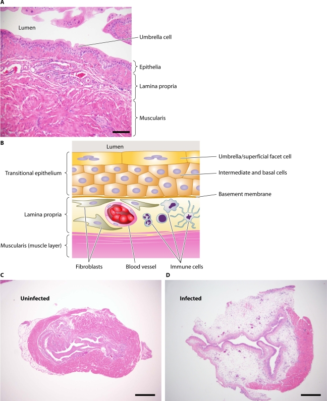
FIG. 1.
Histological and schematic views of the murine bladder. (A) Hematoxylin and eosin (H&E)-stained section from a healthy wild-type C57BL/6 female mouse. Magnification, ×200. Scale bar, 100 μm. (B) Schematic representation of bladder physiology shown in panel A. Umbrella cells line the luminal surface of the transitional epithelium. The basal side of the umbrella cell layer consists of intermediate and basal cells, followed by the lamina propria, the primary site of edema and inflammation during UTI. (C and D) H&E-stained sections from wild-type C57BL/6 mice that were either left untreated (C) or infected for 48 h (D). Note the severe inflammation and edema in the lamina propria of the infected animal. Magnification, ×40. Scale bar, 500 μm.

