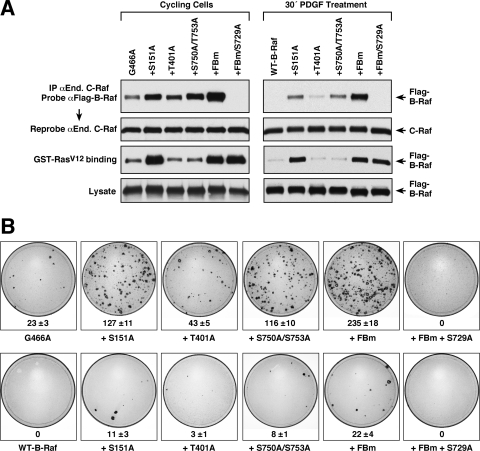
FIG. 6.
Analysis of individual sites of B-Raf feedback phosphorylation. (A) Lysates were prepared either from cycling cells or from PDGF-treated cells expressing the indicated Flag-tagged B-Raf proteins. The lysates were incubated with agarose beads containing GST-RasV12, and the binding of Flag-B-Raf proteins to GST-RasV12 was determined by immunoblot analysis. Endogenous C-Raf proteins were also immunoprecipitated from the lysates and examined for Flag-B-Raf binding. Blots were reprobed to confirm equivalent C-Raf levels, and lysates were examined for Flag-B-Raf levels. (B) NIH 3T3 cells were infected with recombinant retroviruses expressing the indicated B-Raf proteins, and focus formation was visualized after 2 weeks in culture. Focus plates from a representative experiment are shown. The mean number of foci ± standard deviation from at least 3 independent experiments is given below each plate.

