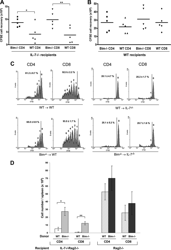
FIG. 3.
Loss of Bim improves T-cell survival but not proliferation in IL-7-deficient hosts. (A) T cells were isolated from LNs of Bim−/− or WT mice, labeled with CFSE, and then transferred intravenously into irradiated IL-7−/− mice. Six days later, spleen cells were stained for CD4 and CD8. Donor T cells were analyzed for CFSE-positive cells by gating on CD4 and CD8. Numbers of CFSE-positive cells in different groups were determined for each spleen. Data were analyzed using CellQuest software (BD Biosciences). Means ± standard deviations for 2 or 3 mice from two independent experiments are shown (*, P = 0.008; **, P ≤ 0.001). (B) CFSE-positive cells were recovered from WT recipient spleens after 6 days of adoptive transfer of T cells. (C) Six days after transfer, T cells were recovered from the spleens of IL-7−/− or WT recipient mice. Donor T-cell homeostatic proliferation profiles were analyzed for CFSE intensity by gating on CD4 or CD8 using flow cytometry. Numbers above the peaks indicate the number of cell divisions. Vertical lines represent the gate used for calculating the percentage of cells that had divided. (D) T cells from Bim−/− or WT mice were transferred to irradiated Rag2−/− or IL-7−/− Rag2−/− recipients. Eight weeks later, spleens were harvested, and CD4 or CD8 T-cell recovery was determined by staining with anti-CD4 and anti-CD8.

