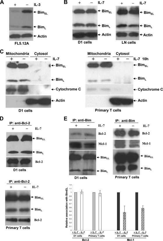
FIG. 5.
IL-7 does not alter Bim synthesis or mitochondrial translocation. Cells were placed in culture for 12 h with or without IL-7. (A) IL-3 was withdrawn from FL5.12A cells for 12 h. Total-cell extracts were subjected to immunoblotting with an antibody against Bim. (B) Total-cell extracts from D1 or LN cells were resolved by SDS-PAGE and subjected to immunoblotting with anti-Bim. (C) Primary T cells were isolated from spleens and LNs of 5 WT mice by negative selection with magnetic beads and were placed in culture for 10 h with or without IL-7. D1 or primary T cells were fractionated into cytoplasmic and mitochondrial fractions, and Bim protein levels were visualized by probing blots with an antibody specific for Bim. Cytochrome c oxidase and actin are shown as markers for mitochondrial and cytosolic proteins, respectively. (D) Cell extracts were prepared from D1 or primary T cells, immunoprecipitated (IP) with anti-Bcl-2, and blotted with anti-Bim or anti-Bcl-2. (E) Total proteins extracted from D1 or primary T cells were subjected to immunoprecipitation with anti-Bim, followed by blotting with anti-Bcl-2, anti-Mcl-1, and anti-Bim. The relative association of Bcl-2 or Mcl-1 with BimEL was quantified using an ImageJ program by measuring densitometry after normalization against BimEL protein.

