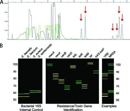
FIG. 1.
(A) Capillary electrophoresis (CE) trace for the antibiotic resistance, and toxin PCR-LDR assay of a vancomycin-resistant E. faecium isolate. The x axis shows the LDR product size, and the y axis shows fluorescence intensity of the bands. Red arrows indicate signals that are unique to the 16S rRNA genes of E. faecium and the vanA gene. (B) The CE data are displayed as a reconstructed gel image generated by a software program developed in our laboratory. The left panel represents the size and fluorescence of the 16S rRNA gene LDR products which act as a positive control and differentiate between the bacteria E. faecalis, E. faecium, and Staphylococcus species. The middle panel represents the LDR signals that identify the presence of methicillin resistance (mecA), vancomycin resistance (vanA, vanB, vanD), tetracycline resistance (tetL tetK, tetM), the toxic shock toxin gene (tst), and the PVL toxin genes (lukS-lukF). Data in the right panel represent two blood cultures. In one, the 16S rRNA gene control identified E. faecium, and the vanA gene was detected, indicating a VRE; in the other the 16S rRNA gene control identified S. aureus, and the mecA and lukS-lukF genes were detected, indicating an MRSA gene encoding the PVL toxin.

