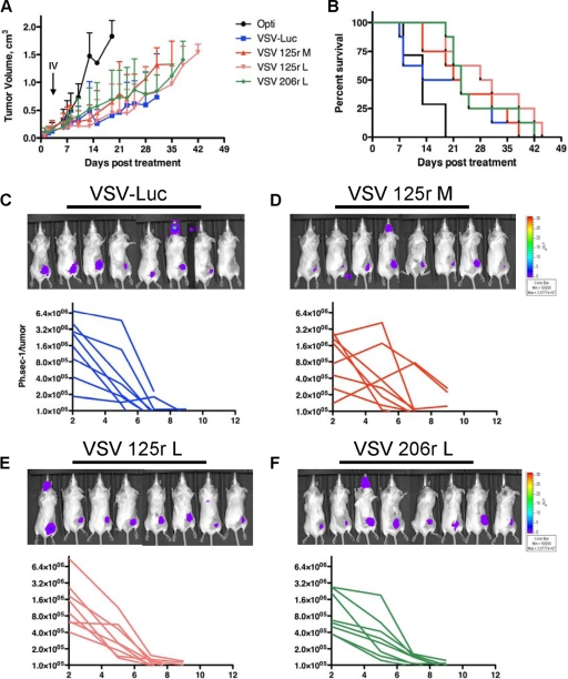
FIG. 7.
MicroRNA-targeted VSVs have equivalent antitumor activities in CT-26 model. (A) CT-26 cells (5 × 10⁶) were injected subcutaneously into the right flank of BALB/c mice, and mice were administered one intratumoral dose of virus (1 × 10⁹) on day 0 and one intravenous dose (1 × 10⁹) on day 3 and monitored for tumor progression. (B) Kaplan-Meier survival curves for the mice in panel A. Images show CT-26 tumor-bearing mice treated with rVSV on day 2, and graphs show the quantification of bioluminescence output per flank, plotted over time, as an indication of viral gene expression for mice treated with VSV-Luc (C), VSV 125r M (D), VSV 125r L (E), and VSV 206r L (F).

