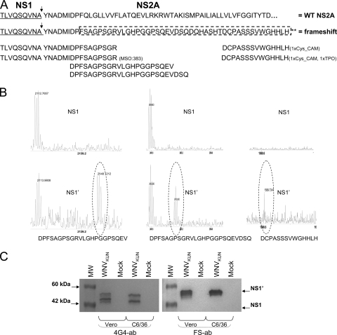
FIG. 2.
Mass spectrometry analysis of NS1 and NS1′ proteins shows that NS1′ is the product of −1 ribosomal frameshifting. (A) Amino acid sequence of the C-terminal region of NS1 (underlined) and N-terminal region of NS2A with the potential frameshift sequence. Arrows indicate cleavage between the NS1 and NS2A proteins. Frameshift amino acids are enclosed in the dashed box, and the sequences of frameshift peptides detected by mass spectrometry are indicated after trypsin and AspN digestion. (B) Peaks of three of the frameshift peptides detected after AspN digestion of NS1′ (bottom panels) but not of NS1 (top panels). (C) Western blot detection with NS1′ frameshift peptide-specific (FS ab) and 4G4 antibodies. Lysates from Vero and C6/36 cells harvested at 3 and 5 days after WNV infection were separated by 10% PAGE, transferred onto nitrocellulose membranes, and incubated with 4G4 (NS1/NS1′ specific) or FS ab (NS1′ specific).

