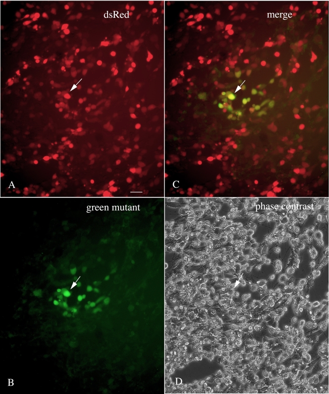
FIG. 2.
VSV-1′DsRed was used to infect a culture of BHK-21 cells with no agarose overlay. Twenty-four hours later, among a population of infected red fluorescent cells (under green light excitation) (A), infected cells that also showed a green color (under blue light excitation) were found (B). Many cells showed both red and green color (see one such cell at the arrow in all four panels) (C). A phase-contrast image of the same field is shown in panel D. Bar, 30 μm.

