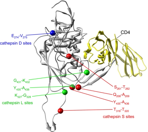
FIG. 4.
Locations of cathepsin L, S, and D cleavage sites on three-dimensional structure of gp120 bound to CD4. The locations of cathepsin cleavage sites were located on the three-dimensional structure of gp120 based on the structure of a gp120 fragment complexed with CD4, described by Huang et al. (47). Cathepsin L sites are indicated in green, cathepsin S sites are in red, and cathepsin D sites are in blue. The structure of CD4 is shown in yellow. Numbering is based on the sequence of MN-rgp120.

