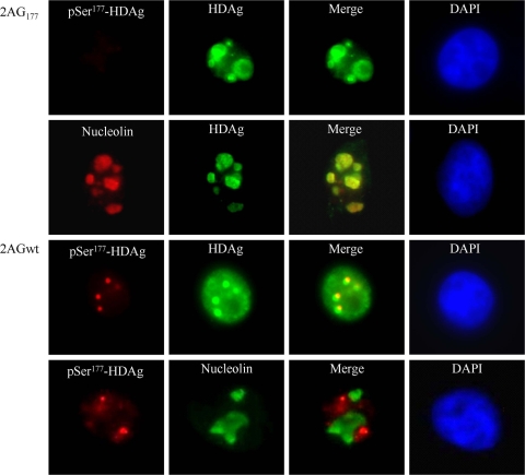
FIG. 1.
Localization of Ser177-phosphorylated HDAg was restricted to nuclear bodies, while its unphosphorylated counterpart accumulated predominantly in nucleoli. 293T cells were transfected either with an HDV replication-competent construct, 2AGwt, or with an HDV antigenomic RNA replication-deficient construct, 2AG177. At 2 days posttransfection, cells were dually immunostained with antibodies against HDAg, Ser177-phosphorylated HDAg (pSer177-HDAg), or nucleolin and were counterstained with 4′,6-diamidino-2-phenylindol (DAPI). pSer177-HDAg is indicated in red, HDAg in green, and DAPI counterstaining in blue.

