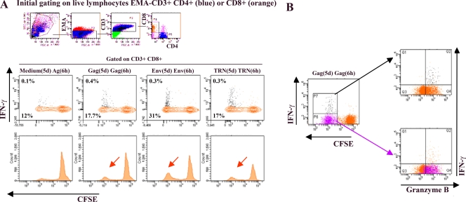
FIG. 2.
Polyfunctional HIV-specific CD8+ T cells in vaccinated macaque AL49 at 28 weeks p.i. PBMCs from macaque AL49 were labeled with CFSE and cultured in the presence of 2 μg/ml of specific pools of HIV peptides Gag, Env, TRN, or medium only for 5 days [designated Gag(5d), Env(5d), TRN(5d), and medium(5d)]. On day 5, cells were harvested and restimulated for 6 h with the same mix of peptides [Ag(6h)] in the presence of costimulatory antibodies and brefeldin A. Cells then were surface stained with anti-CD3, anti-CD8, and anti-CD4 MAbs in the presence of EMA (to allow the exclusion of dead cells) and subsequently permeabilized and stained with anti-IFN-γ and anti-granzyme B MAbs. For flow cytometry analysis, we gated on low-FSC/SSC, EMA−, CD3+, and large CD8+ T-cell populations (colored in orange). The proportions of cells producing IFN-γ (contour plot, upper number) and proliferating (CFSE dilution; contour plot, lower number; and histogram [arrow]) and dot plots (B) in response to specific antigens are presented. Frequencies for antigen-specific responses are reported (see Results) as the percent cytokine-secreting or proliferating CD8+ T cells after the subtraction of backgrounds obtained with cells cultured for 5 days with medium only and restimulated for 6 h with relevant mixes of peptides [medium(5d) Ag(6 h)]. (B) Proliferating-only (purple dots) as well as proliferating and IFN-γ-producing (black dots) Gag-specific CD8+ T cells are superposed to the total CD8+ T-cell population (in orange) for granzyme B expression detection (left panels). For simplicity, only the Gag response is shown, but Env and TRN antigens gave similar results.

