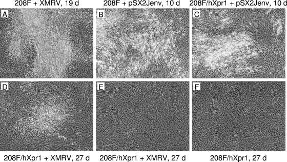
FIG. 1.
Morphology of transformed foci in 208F fibroblasts. 208F and 208F/LhXpr1SN cells seeded the day before at 1 × 10⁵ to 2 × 10⁵ cells per 6-cm dish were infected with XMRV+LAPSN virus (produced by HTX/LAPSN cells infected with virus from 22Rv1 cells) (A, D, E), were transfected with the pSX2Jenv plasmid by using the calcium phosphate method (B, C), or were treated with culture medium only (F). The cells were trypsinized and replated in multiple 6-cm dishes 1 day (A, D to F) or 10 days (B, C) after treatment. Areas with (A to D) and without (E and F) foci are shown. Cell layers were photographed at the indicated times after replating. d, days.

