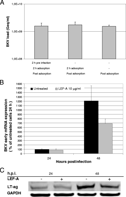
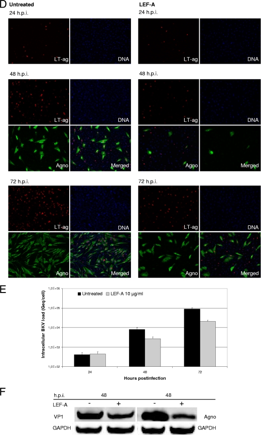
FIG. 2.
Influence of LEF-A on different steps in the BKV life cycle. RPTECs were seeded and infected as described earlier. (A) The influence of LEF-A on BKV adsorption and entry was monitored by comparing LEF-A addition 2 h before, together with, or 2 h after BKV infection. Supernatants were harvested at 72 h.p.i., and extracellular BKV loads were measured by qPCR as described. (B) LT-ag transcription was measured 24 and 48 h.p.i. Total RNA was extracted using the mirVana Paris kit (Ambion, Applied Biosystems) and treated with DNase turbo (Ambion, Applied Biosystems) before cDNA was generated from 225 ng RNA per sample using the High Capacity cDNA kit (Applied Biosystems). LT-ag transcripts were quantified by RT-qPCR and normalized to the levels of endogenous human hypoxanthine phosphoribosyltransferase (huHPRT) transcripts by the 2−ΔΔC(T) method (5, 28). Results are presented as the changes in LT-ag transcript levels, with the level in the untreated sample 24 h.p.i. arbitrarily set to 100%. (C) LT-ag protein levels at 24 and 48 h.p.i. were examined by Western blotting. RPTECs were lysed in cell disruption buffer (mirVana Paris kit; Ambion), and Western blotting was performed as described previously (5) using polyclonal rabbit anti-LT-ag serum (20) and a monoclonal antibody directed against the housekeeping protein glyceraldehyde-3-phosphate dehydrogenase (GAPDH) (Abcam). The secondary antibodies used were IRDye800CW goat anti-rabbit IgG (Rockland) and Alexa Fluor 680 goat anti-mouse IgG (Invitrogen). (D) Early and late protein expression was investigated by indirect immunofluorescence staining. RPTECs were methanol fixed 24, 48, and 72 h.p.i., blocked with 3% goat serum in phosphate-buffered saline (PBS) for 30 min, and then treated as described earlier (27). The primary antibodies, SV40 LT-ag monoclonal (Pab416; Calbiochem) (red) and polyclonal rabbit anti-agnoprotein serum (20) (green), and the secondary antibodies, Alexa fluor 568 goat antimouse (Invitrogen) and Alexa fluor 488 goat antirabbit (Invitrogen), were used. Cell nuclei (blue) were stained with DRAQ5 (Biostatus). Images were collected using a Nikon TE2000 microscope equipped and processed with the NIS Elements Basic Research software program, version 2.2 (Nikon Corporation). (E) Intracellular BKV DNA loads were quantified from RPTECs harvested at 24, 48, and 72 h.p.i. After DNA extraction, BKV DNA loads were measured by qPCR and normalized for cellular DNA using the aspartoacylase (ACY) qPCR (5, 35, 36). Data are presented as Geq/cell. (F) Late protein expression was detected by Western blotting 48 h.p.i. as described above (5), using as primary antibodies polyclonal rabbit anti-VP1 serum (17), polyclonal rabbit anti-agnoprotein serum (20), and the monoclonal antibody directed against GAPDH (Abcam).


