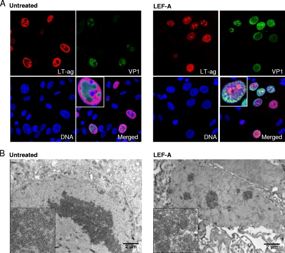
FIG. 3.
Influence of LEF-A on nuclear BKV replication architecture. (A) For confocal microscopy, RPTECs were seeded in fibronectin-coated chamber slides, infected, and treated with LEF-A (10 μg/ml) as described earlier. At 72 h.p.i., cells were fixed in 4% paraformaldehyde, followed by methanol permabilization and blocking with 3% goat serum in PBS for 30 min. Then, indirect immunofluorescence was performed as described earlier (27) using as primary antibodies an SV40 LT-ag monoclonal (Pab416; Calbiochem) (red) and polyclonal rabbit anti-VP1 serum (17) (green) and the secondary antibodies Alexa fluor 568 goat anti-mouse IgG (Invitrogen) and Alexa fluor 488 goat anti-rabbit IgG (Invitrogen). Cell nuclei (blue) were stained with DRAQ5 (Biostatus). Confocal microscopy analysis was performed using a microscope (Axiovert 200; Carl Zeis, Inc.) equipped with an LSM510 confocal module and processed using the LSM5 software program, version 3.2 (Carl Zeiss, Inc.). (B) For IEM analysis, RPTECs were grown in 6 wells, infected, and treated with LEF-A (10 μg/ml) as earlier described. At 48 h.p.i., cells were fixed with 4% formaldehyde, washed in 0.12% glycin, scraped off, and pelleted in 12% gelatin. The pellet was placed in 2.3 M sucrose overnight, cut in cubes, mounted on cryo-pins, frozen by immersion in liquid nitrogen, and sectioned using a Leica EM UC6 ultramicrotome. The sections were submerged in 1% cold-water fish skin gelatin overnight and incubated with polyclonal rabbit anti-VP1 serum (17) and then with protein A-gold (10 nm). The specimens were contrasted with a mixture of uranyl acetate and methylcellulose and examined by using a Jeol 1010 transmission electron microscope.

