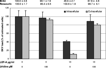
FIG. 4.
Impact of uridine on BKV replication and cellular viability in LEF-A-treated RPTECs. RPTECs were seeded and infected as earlier described. At 2 h.p.i., virus was removed and replaced with LEF-A-containing medium with or without exogenous uridine at 100 μM. For intracellular DNA measurements, cells were harvested 48 h.p.i., and for extracellular BKV load, supernatants were harvested 72 h.p.i. BKV and cellular DNA loads were measured by qPCR as described earlier, and BKV loads of untreated cells were set as 100%. In addition, RPTEC cellular DNA replication was measured by cell proliferation ELISA, BrdU, and total cellular metabolic activity determined using TOX-8 at 48 h.p.i. as described earlier. Absorbance for untreated cells was set as 100%.

