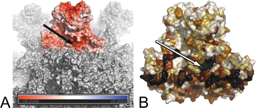
FIG. 3.
A conserved groove in birnavirus VP2 proteins. (A) Surface representation of the IPNV VP2 SVP. One trimer is colored according to the electrostatic potential on the solvent-accessible surface as calculated by the Adaptive Poisson-Boltzmann Solver (2) and displayed in PyMo (http://www.pymol.org/). Two negatively charged patches are obvious, one at the center of the trimer tip and the other at the conserved groove at the base of the trimer (arrow). (B) Representation of the sequence conservation across birnaviruses mapped onto the molecular surface of a VP2 trimer (black-brown-white gradient from most to least conserved residues). As in panel A, the arrow points to the conserved groove.

