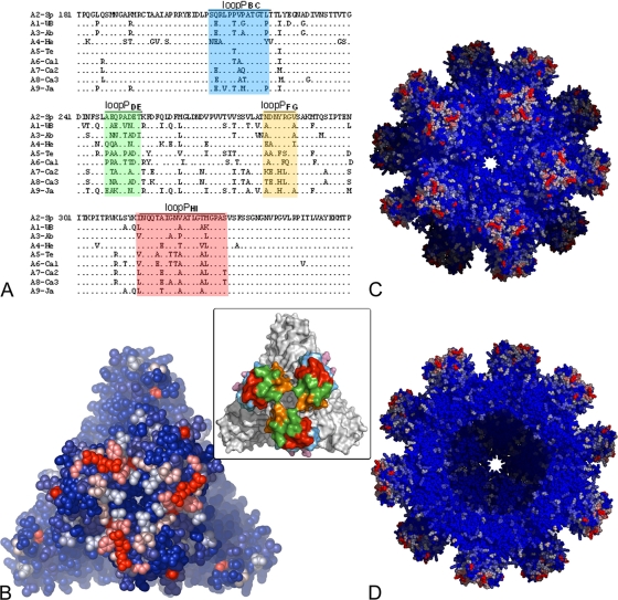
FIG. 6.
Sequence variability between IPNV serotypes. (A) Sequence alignment of domain P from the nine IPNV serotypes. The variable loops are colored according to the inset of Fig. 5 (shown also in panel B). (B) The IPNV VP2 trimer in an all-atom representation (as spheres). (C and D) The SVP viewed along a 5-fold axis. A blue-white-red color gradient was used for strictly conserved residues to highly variable ones as estimated by the ConSurf server. In panel D, five trimers from the front part of the particle were removed to reveal the internal icosahedral shell.

