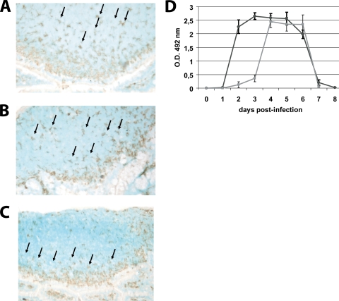
FIG. 2.
UV-treated virus did not induce the migration of DCs to the SED of PPs. PP from jejunum of RV EDIMwt-infected mice or from mice inoculated with UV-treated virus were obtained and cryopreserved at 0 (control mice) and 48 h p.i. The frozen sections of the PPs were stained with a CD11c-specific hamster MAb (clone N418) and developed with a secondary goat anti-hamster IgG Ab coupled to peroxidase using 3-amino-9-ethylcarbazole chromogen substrate. (A) Histological analysis of CD11c+ cells in PPs of noninfected mice. (B) CD11c+ cells in PPs at 48 h p.i. with UV-treated virus. (C) CD11c+ cells in PPs at 48 h p.i. with infectious RV. Arrows indicate CD11c+ cells. For panels A, B, and C a representative picture from 30 PPs is shown. (D) RV shedding curves. Groups of four mice were inoculated orally with 104 FFU of murine RV EDIMwt (dark gray line) or UV-treated RV EDIMwt. (light gray line). Fecal rotavirus antigen was measured from day 0 to 8 by ELISA, and the results were expressed as the net optical density (O.D.) value. Bars represent the SD. The data shown are representative of three independent experiments.

