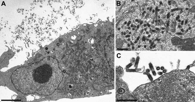
FIG. 5.
Accumulation of intracellular viruslike structures in SAD RABV M-infected cells is comparable to the authentic SAD L16 virus. Electron microscopic analysis of SAD RABV M-infected BSR T7/5 monolayer cell cultures. (A) Intracellular viruslike structures. Scale bar, 4 μm. (B) Higher magnification of intracisternal structures indicate accumulation in the rER. Scale bar, 500 nm. (C) Extracellular virions derived from budding at the plasma membrane. Scale bar, 500 nm.

