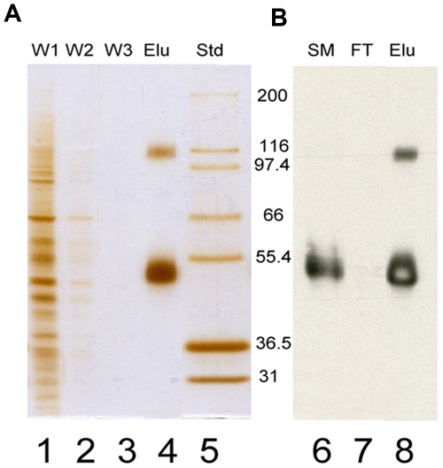
Figure 1. Quality control of RhCG purification.
A 10% SDS PAGE was divided in two parts (A and B) after migration. A, Silver staining. lanes 1 to 3: aliquots of the three Washes (W1, W2, W3); lane 4: HA peptide Eluted fraction (Elu); lane 5: molecular weight Standards (Std) from Invitrogen. B, Western blot probed with an anti HA and revealed with Amersham ECL Western blotting detection reagent (GE Healthcare, Buckinghamshire, UK), RhCG can be detected in starting material (SM) on lane 6 and in the HA peptide Eluted fraction (Elu) on line 8 but not in the flow through (FT) on lane 7. Arrows indicate the two bands of 50 kDa and 110 kDa detected by both silver staining and western blot analysis, most likely corresponding to monomeric and oligomeric forms of RhCG.

