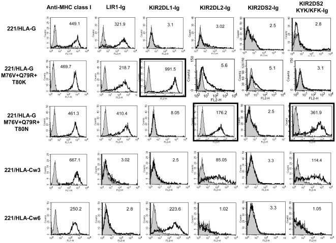
Figure 3. 221/HLA-G mutated in three contact residues are recognized by KIR-Ig fusion proteins.
Wild-type, triple mutated 221/HLA-G, 221/HLA-Cw3 and 221/HLA-Cw6 were stained with various fusion proteins followed by secondary antibody staining. Gray histograms represent background secondary antibody staining. For confirmation of expression level, cells were stained with anti-MHC class I mAb (left panel). Black frames emphasize the unique KIR-Ig binding to the 221/HLA-G triple mutants. Shown is a representative experiment of at least three independent experiments.

