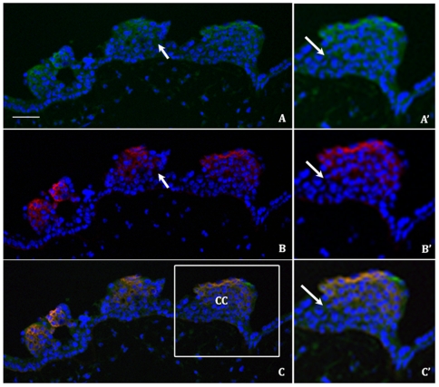
Figure 6. The conformed and FHC forms of HLA-G are differentially expressed in trophoblast cell columns.
Double immunofluorescent staining of representative first trimester placental sections (week 10–11) with antibody MEM-G9 (A,A′) and 4H84 (B,B′) and the combined image (C,C′) (scale bar 50 µM). DAPI (blue), conformed HLA-G (green) and FHC HLA-G (red) staining is observed in the panels with the magnified inset (boxed area in image C) viewed in panels A′,B′,C′. Several areas where differential expression of conformed and FHC HLA-G are indicated by arrows. CC indicates a trophoblast cell column.

