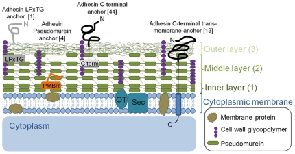
Figure 4. Putative cell envelope topography of M1.
Ultrastructural studies of M1 [88], [89] show that the cell wall is composed of three layers and is comparable to the organization seen in Gram positive bacteria [90]. The three layers can be described as: (1) a thin electron-dense inner layer composed of compacted newly synthesised pseudomurein, (2) a thicker less-electron-dense middle layer which is also composed of pseudomurein, and (3) a rough irregular outer layer that is distal to the pseudomurein layers and assumed to be composed of cell wall glycopolymers, wall-associated proteins and possibly other components. Representative adhesin-like proteins with different cell-anchoring domains are shown. The number of these proteins predicted in the M1 genome is shown in brackets. OT, oligosaccharyl transferase; Sec, Sec protein secretion pathway; PMBR, pseudomurein binding repeat (PF09373); M1-C, M1 adhesin-like protein conserved C-terminal domain.

