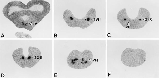
Figure 3.
X-ray autoradiographs showing the distribution of prepro-UII mRNA in the frog brainstem and spinal cord. Coronal sections were hybridized with the antisense (A–E) or the sense (F) prepro-UII riboprobe and were exposed for 2 weeks onto x-ray film. IV, trochlear nucleus; VI, abducens nucleus; VII, facial nucleus; IX, glossopharyngeal nucleus; XII, hypoglossal nucleus; VH, ventral horn of the spinal cord.

