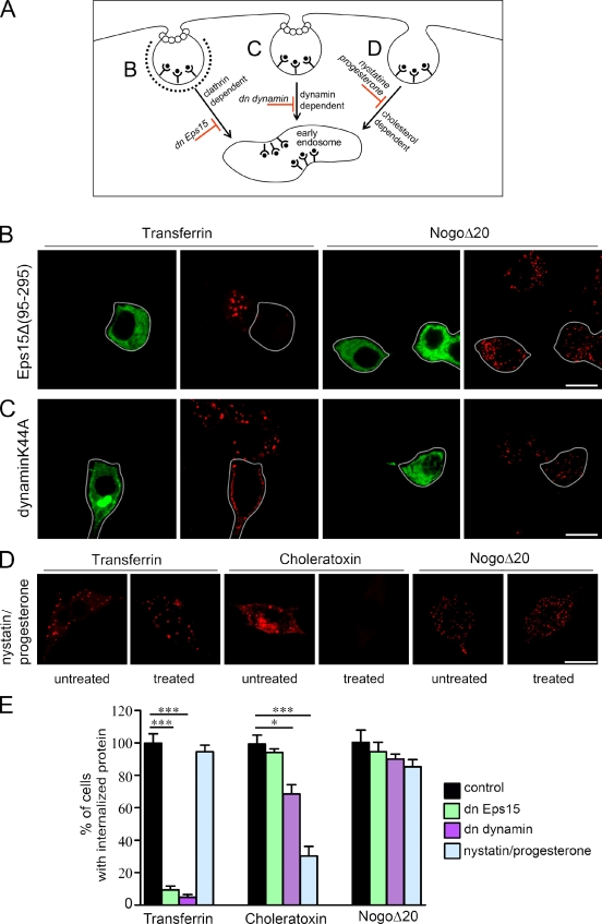
Figure 2.
Internalization of NogoΔ20 occurs independently of Epsin15, dynamin II, and cholesterol. (A) Schematic representation of different endocytotic pathways and their blockers. (B and C) PC12 cells were transfected with GFP-tagged Eps15Δ (95–295) (B, green), or GFP-dynIIK44A (C, green). 24 h later, cells were incubated with 300 nM NogoΔ20-T7 (red) or 100 nM transferrin-biotin (red) for 30 min at 37°C. In Eps15Δ (95–295) and dynIIK44A-transfected cells, transferrin uptake was blocked, but uptake of NogoΔ20 was not. White lines indicate the outlines of GFP-expressing cells. Bars, 10 µm. (D) PC12 cells were either left untreated or were pretreated with nystatin and progesterone overnight, and then incubated with transferrin, choleratoxin, or NogoΔ20-T7 for 30 min at 37°C in the absence or presence of drugs. Although the internalization of choleratoxin was inhibited (red), the internalization of transferrin (red) and NogoΔ20 (red) was not affected. Representative optical sections from three independent experiments are shown. Bar, 10 µm. (E) Quantification of protein uptake after various cell treatments. The percentage of cells with internalized protein is given as the mean ± SEM (*, P < 0.05; ***, P < 0.001; Student’s t test).

