Figure 6.
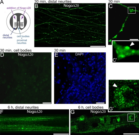
Upon internalization, NogoΔ20 is retrogradely transported from the neurites to the cell bodies of dissociated DRG neurons. (A) Schematic representation of a compartmentalized chamber. (B–F) Representative immunofluorescence images of retrogradely transported NogoΔ20 (300 nM) at indicated time points in dissociated DRG neurons that were cultured in compartmentalized Campenot chambers. (B) Uptake of NogoΔ20 (green) in the distal neurites at 30 min of incubation. Bar, 20 µm. (C) NogoΔ20-positive vesicles (green, arrowhead) in the distal neurites. Bar, 10 µm. (D) NogoΔ20 could not be observed in the cell body compartment 30 min after NogoΔ20 addition. (E) Cell bodies were stained with DAPI (blue). Bar, 40 µm. (F and G) NogoΔ20-positive distal neurites (F) and cell bodies (G) 6 h after addition of NogoΔ20 to the distal compartment. The arrowhead indicates NogoΔ20-positive vesicles in the cell body. The inset panels in C and G show enlarged views of the boxed regions. Bars, 20 µm.

