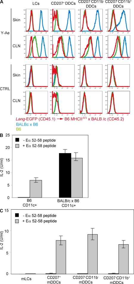
Figure 6.
DDCs do not present Eα52-68 LC-derived peptides. (A) Flow cytometry analysis of LCs, CD207+, CD207− CD11b−, and CD207− CD11b+ DDCs isolated from Langerin-EGFP→B6 MHCIIΔ/Δ x BALB/c chimeras (red) and from B6 (green) and B6 x BALB/c (blue) mice before (skin) and after (CLN) their migration to CLN. Cells were separately stained with Y-Ae and isotype control staining (CTRL). (B) CD11c+ DCs isolated from B6 and B6 x BALB/c mice were cultured together with H30 T cells in the presence or absence of 3 µg/ml Eα52-68 peptide. The content of IL-2 present in supernatant was determined after 24 h of culture. (C) LCs, CD207+, CD207− CD11b−, and CD207− CD11b+ DDCs isolated from the CLNs of Langerin-EGFP→B6 MHCIIΔ/Δ x BALB/c chimeras were cultured together with H30 T cells that are specific for Eα52-68–I-Ab complexes and in the presence or absence of 3 µg/ml Eα52-68 peptide. The content of IL-2 present in supernatant was determined after 24 h of culture. Data shown are representative of three independent experiments. Error bars correspond to SEM.

