Figure 1.
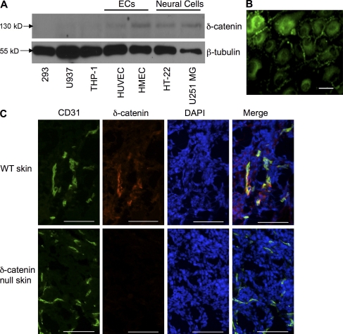
δ-Catenin is expressed in vascular endothelium. (A) Protein lysates collected from neuronal cells (HT-22 and U251 MG), endothelial cells (ECs; HUVEC and HMEC), epithelial cells (293), and leukocytes (THP-1 and U937) were subjected to Western blot analysis and probed with a δ-catenin–specific antibody. (B) β-tubulin was used as a loading control. Immunofluorescent staining for δ-catenin was performed in cultured HUVECs. (C) Mouse skin tissue sections from wild-type and δ-catenin–null mice were analyzed by immunofluorescent double staining using antibodies against CD31 for endothelium, and δ-catenin. Nuclei were stained with DAPI. 400× magnification. Each experiment was repeated three times, and representative images were shown. Bar, 200 µm.

