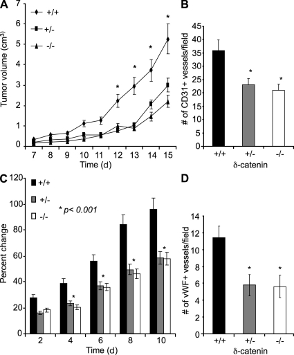
Figure 5.
Genetic deletion of δ-catenin in mice impairs pathological angiogenesis in a dose-dependent manner. 3LL tumor cells were injected subcutaneously into sex- and age-matched syngeneic δ-catenin homozygous-null, heterozygous-null, and wild-type littermates. (A) Tumor growth was measured by caliper for 15 d, and tumor volume was calculated and plotted (n = 10 mice/group; *, P < 0.001). (B) Tumor tissue sections from each group were analyzed by CD31 immunofluorescent staining, and CD31 positive vessels were counted from 10 randomly selected high-power fields under microscopy. Mean and SE were plotted. *, P < 0.05. (C) 6-mm biopsy punches were made in wild-type littermates, δ-catenin heterozygous-null, and δ-catenin homozygous-null mice. Wound size was measured daily. The percent changes of wound size over the original size were plotted (n = 7 mice/group; *, P < 0.01). (D) The vascular density measurement by vWF staining was performed in wounded tissues on day 3. Mean and SE were plotted. *, P < 0.01.

