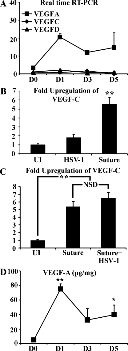
Figure 4.
VEGF-A expression is up-regulated after HSV-1 infection. (A) Corneal levels of VEGF-A/C/D were quantified by real-time RT-PCR and normalized per 1,000 transcripts of the housekeeping gene β-actin with values given at the indicated day PI. Although HSV-1 infection induced up-regulation of VEGF-A, no up-regulation of VEGF-C or -D was observed. **, P < 0.01, comparing infected to uninfected groups. The figure is a summary of two experiments. n = 3 per group per experiment. (B) However, at day 5 after corneal suture placement VEGF-C messenger RNA was significantly up-regulated relative to both untreated and HSV-1–infected corneas, here expressed as fold induction using the housekeeping gene β-actin for normalization. **, P < 0.01 comparing the suture-induced VEGF-C level to the HSV-1 and uninfected (UI) groups. The figure is a summary of two experiments, n = 3 per group per experiment. (C) To determine if HSV-1 infection may suppress VEGF-C expression, sutured mice were either mock infected or infected with HSV-1 at the time of suture placement and the corneas were collected at day 5 after suture/PI. HSV-1 infection did not affect VEGF-C expression after suture placement. A summary of two experiments is shown with a total of n = 6 per group. **, P<.01 comparing the uninfected to the other two groups. NSD, no significant difference. (D) Protein levels for VEGF-A were measured by cytokine bead array after HSV-1 infection, expressed as picograms of VEGF-A per milligram of wet cornea mass. VEGF-A protein expression was rapidly up-regulated and remained up-regulated throughout the course of the study. **, P < 0.01; *, P < 0.05, comparing levels of VEGF-A protein at time points PI to uninfected (day 0 [D0]) control. The figure is a summary of two experiments, n = 6 corneas per time point. Error bars in A–D represent the SEM based on the results of each cornea sample summarized for both experiments.

