Figure 1.
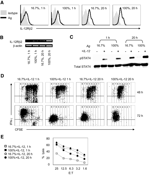
High amount of antigen stimulation increases expression of IL-12Rβ2 and promotes IL-12-mediated CD8+ effector. Naive OT-I T cells stimulated with varying ratios of antigen-expressing fibroblasts (BOK) versus nonantigen-expressing fibroblasts (MEC II); 1:5 (16.7%) or 1:0 (100%) for 1 h or 20 h. (A) Surface expression of IL-12Rβ2 on CD8+ gated OT-I T cells at 48 h. (B) RT-PCR of IL-12β2 in CD8+ selected OT-I T cells at 24 h. The β-actin mRNA serves as an internal control. (C) OT-I T cells were stimulated for 1 h or 20 h with 16.7% or 100% ratio of BOK/MEC II cells in the presence or absence of IL-12 (2 ng/ml). Cells were harvested and lysed and protein subjected to Western blot analyzed for STAT4 [phosphorylation (pSTAT4) and total] with specific antibodies, respectively. (D and E) Naive CFSE-labeled OT-I CD8+ T cells stimulated with 16.7% or 100% ratio of BOK/MEC II cells for 1 h or 20 h in the presence of IL-12 (2 ng/ml) were subjected to ICS for IFN-γ and flow cytometry analysis at 48 h and 72 h (D). The OT-I cells were harvested at 72 h and evaluated for cytolylic ability in a standard 4-h chromium release assay (E). The results are representative of three independent experiments.

