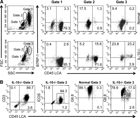
Figure 5.
The B7RP-1 ICOS ligand is up-regulated on nonhematopoietic cells and leukocytes in the intestinal epithelium of IL-10−/− mice. (A) Freshly isolated cells consisting of cIELs and enterocytes from the colonic epithelium of normal BALB/c and IL-10−/− mice were stained for expression of CD45 LCA and B7RP-1. Cells were defined as hematopoietic cells based on CD45 LCA expression or nonhematopoietic cells based on a lack of CD45 LCA expression. Three populations (Gates 1, 2, and 3) in cell isolates from each animal type were defined by properties of forward or side scatter (FSC/SSC, respectively) by flow cytometry. Two notable differences were observed in normal versus IL-10−/− mice. First, there was a large percent of large granular LCA+ cells in the Gate 2 population of IL-10−/− mice that did not exist in normal animals. Second, approximately one-half of all of the cells in the Gate 3 population of IL-10−/− mice was B7RP-1+ cells, which consisted of roughly equivalent percentages of hematopoietic and nonhematopoietic cells. To characterize the cells in Gate 2, freshly isolated cIELs from IL-10−/− mice were stained for CD3 and ICOS expression. (B) The majority of those cells included CD3+, ICOS+, and LCA+ cells, indicating that they were activated T cells. Additionally, cells from normal and IL-10−/− mice were stained using the G8.8 antibody that reacts with Ep-CAM on epithelial cells and some APCs [31, 32]. Cells from normal mice in Gate 3 were almost entirely G8.8+, LCA– epithelial cells, whereas some cells in Gate 3 from IL-10−/− mice were G8.8+, LCA+ leukocytes that were probably macrophages or DCs. This implies that there is a loss of epithelial cell homeostasis in IL-10−/− with colonic pathology. Data are representative of two to three independent experiments.

