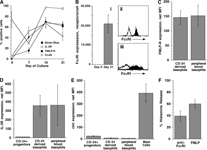
Figure 2.
Phenotypic characterization of CD34-derived basophils. Progenitor cells from five different donors were used for these experiments. (A) Time-course for the appearance of basophil markers as assessed by flow cytometry and Alcian blue staining, represented as the fraction of cells positive for a particular marker; Alcian blue (n=8), FcεRI (n=2), fMLP-R (n=3), and IL-3R (n=2). (B) Expression of FcεRI on CD34-derived basophils (n=10), expressed as the number of receptors (i) and comparison of FcεRI expression on peripheral blood basophils (ii) versus CD34-derived basophils (iii). Solid, Anti-FcεRI; outline, isotype control. The number of FcεRI receptors was calculated based on previous calibration of the flow data to the amount of IgE that could be eluted from the peripheral blood basophils [24]. (C) Expression of fMLP-R on CD34-derived basophils (n=4) versus peripheral blood basophils (n=3). (D) Expression of IL-3R on CD34+ progenitors (n=4) versus CD34-derived basophils (n=4) and peripheral blood basophils (n=15). (E) Expression of c-kit on CD34-derived basophils (n=2) versus CD34+ progenitors (n=3), peripheral blood basophils (n=3), and human lung mast cells (n=4). (F) Histamine release of CD34-derived basophils in response to an optimal concentration of anti-FcεRI antibody (n=4) and fMLP peptide (n=3).

