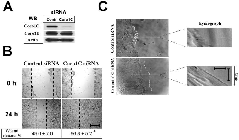
Figure 1. Depletion of Coronin 1C increases motility and modulates lamellipodia dynamics in IECs.

SK-CO15 cells treated with control- or Coronin 1C-specific siRNAs. (A) Cells were lysed and immunobloated for coronins 1C and 1B to ensure efficient and selective Coronin 1C knock-down and for actin to ensure equal protein loading. (B) Representative phase contrast images showing wound closure in control and Coronin 1C-depleted cell monolayers. Bar, 400μm. Wound widths measured 24 hours after wounding and expressed as a % of closure of the initial wound widths at time 0 are indicated under the figure. *, p<0.01 compared to control. (D) Phase contrast images showing the shape of lamellipodia (outlined in white) at the leading edge of the wound 120 minutes after wounding. Kymographs were generated from 180-frame 1 minute interval image sequences using pixel intensities along 1-pixel wide line shown. Vertical bar, 60 seconds; horizontal bar, 20 μm. Note the fuzzy pattern on kymograph reflecting the fast withdrawal rate of the formed protrusions in control cells and smooth pattern representing the persistence of lamellipodial protrusion in Coronin 1C-depleted cells.
