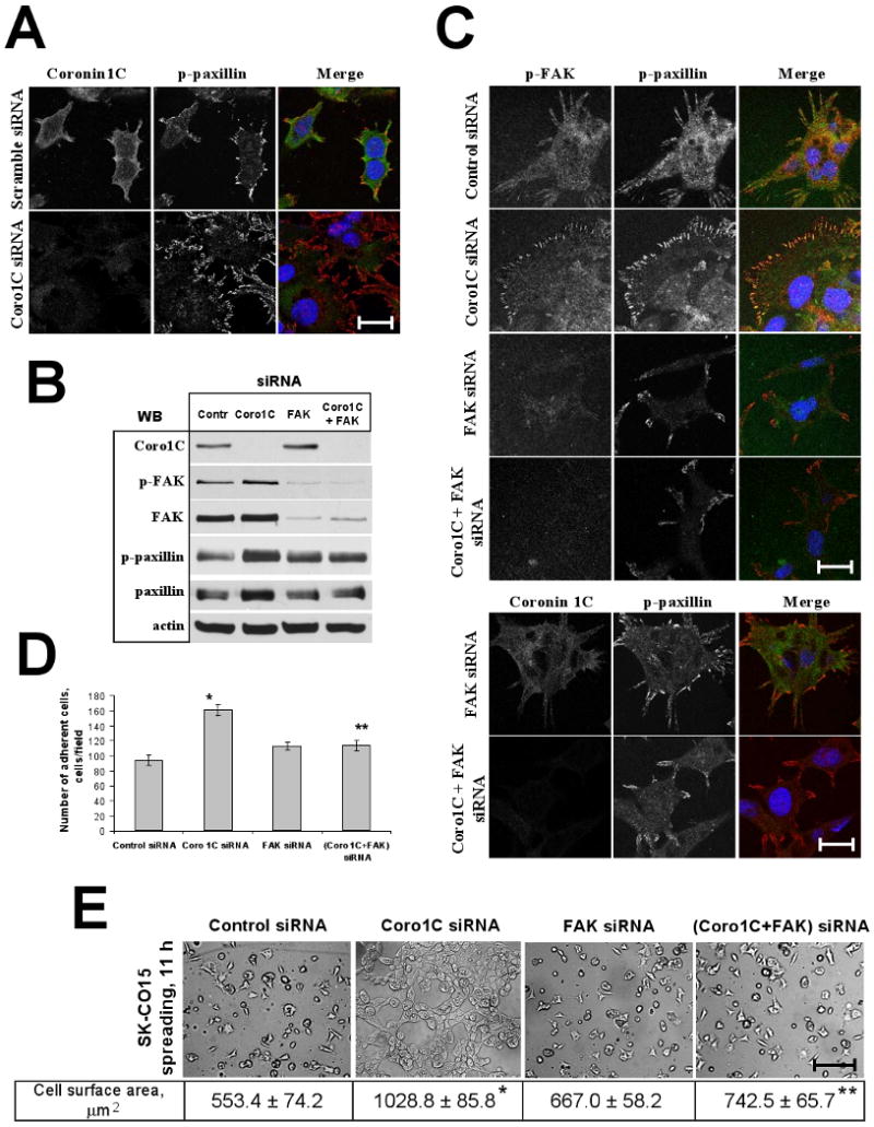
Figure 3. Coronin 1C regulates cell-matrix adhesion through FAK-mediated signaling.

SK-CO15 cells transfected with indicated siRNAs were replated on ECM gel and allowed to spread for 11h. (A) En-face confocal images showing morphology of FAs visualized by immunolabeling for p-paxillin. Note increase in number and density of peripherally located FAs in Coronin 1C-depleted cells. Bar, 25 μm. (B) Cells were lysed and immunoblotted for Coronin 1C, p(397)FAK, FAK, p(118)paxillin, paxillin, and actin (loading control). Note increase in the amount of phosphorylated FAK and paxillin in Coronin 1C-depleted cells (C) En-face confocal images showing localization of phospho-paxillin and phospho-FAK in spreading cells. Note that knock-down of FAK reverses accumulation of peripheral FAs in Coronin 1C-depleted cells. Bar, 25 μm. (D, E) Cell-matrix adhesion and spreading of single and dual Coronin 1C- and FAK knock-downs was analyzed as described in Fig. 2. (D) Average number of adherent cells per microscopic field 3 hours after plaiting. (E) Representative phase contrast image of spreading cells. Bar, 200 μm. Average cell surface area of spreading cells is shown. *, p<0.01 compared to control; ** p<0.05 compared to Coronin 1C siRNA-transfected cells.
