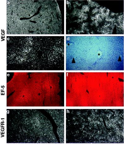
Figure 6.
Localization of VEGF (a–d) and VEGFR-1 (g, h) mRNA in adult liver. (a) Homogenous VEGF staining in normoxic liver. (b) Patchy VEGF expression during hypoxia. (c) Magnification of b, showing intensified VEGF expression around central veins (marked by arrowheads in d) and reduced expression around periportal fields (indicated by asterices in d). (d) Bright-field image of c. (e) Weak EF5 staining in normoxic liver. (f) Hypoxic regions in liver are shown by strong EF5 staining, especially around central veins. (g) Low expression of VEGFR-1 in normoxic liver. (h) Increased VEGFR-1 expression around central veins during hypoxia. Original magnification: ×6.25 (a, b, g, h); ×25 (e, f); ×50 (c, d).

