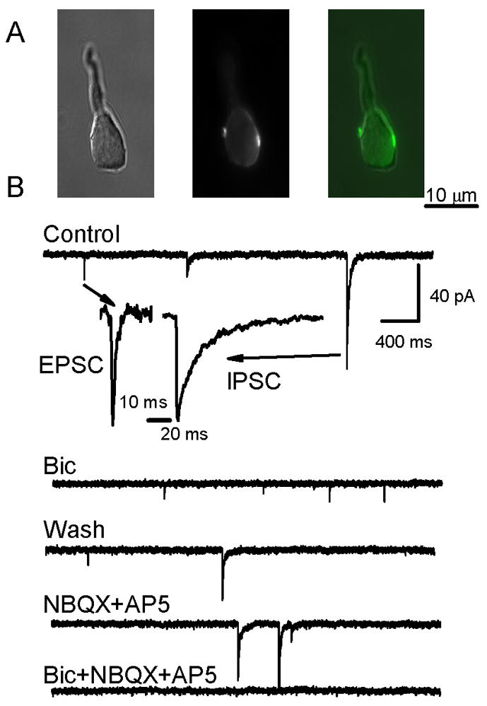
Figure 2.

Retained nerve terminals on acutely isolated NTS neurons are stained with FM1-43 and can be glutamatergic or GABAergic. A. Photomicrographs of a representative neuron (DIC image, left) that showed multiple fluorescent puncta (middle) along the edge of an NTS neuron cell body following loading of FM1-43 following loading using 10 mM external K+ ACSF (center). Superimposition of DIC and epi-fluorescence images (right) shows two terminals on opposite sides of the neuron. B. In another representative isolated NTS neuron, whole cell currents were recorded with nystatin perforated patch electrodes and revealed the presence of two populations of synaptic events, small amplitude, fast spontaneous synaptic currents and larger amplitude, slow decaying synaptic currents. Insets show expanded traces normalized to the same peak current to illustrate that IPSCs had prolonged decay phases, whereas EPSCs rapidly returned to baseline. Note that neurons were superfused via the Y-tube system throughout the experiment at constant flow and Control was normal ACSF. Bicuculline (100 μM) reversibly blocked large-amplitude, long-duration, synaptic currents and identified them as GABAergic IPSCs while preserving small-amplitude, brief synaptic currents (EPSCs). Wash with drug-free control solution restored the spontaneous IPSCs mixed with EPSCs. NBQX (20 μM) and AP5 (100 μM) blocked the fast events identifying them as glutamatergic EPSCs, but sIPSCs remained. All synaptic currents were blocked by combined bicuculline and NBQX.
