
Fig. 9.
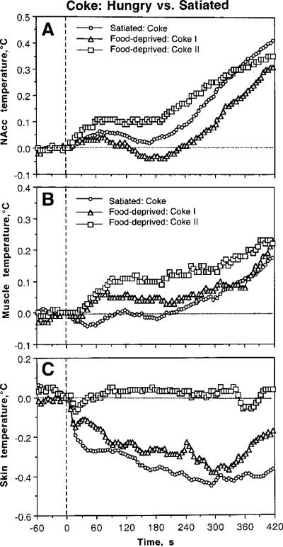
Differences in phasic changes in brain (A), muscle (B), and skin (C) temperatures associated with Coke presentations in food-deprived and satiated conditions. Each graph shows three curves, representing relative temperature changes (5-s bins) after two (I and II) Coke presentations in food-deprived conditions and regular Coke presentation in control, satiated conditions. Hatched lines show zero time and zero temperature change.