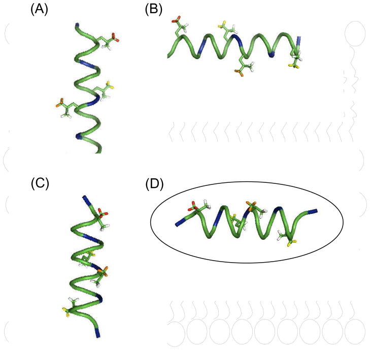
Figure 6.
Model of the possible structures and orientations of KL4 in the lipid bilayer: (A) a canonical α-helix in a transmembrane orientation; (B) an α-helix in the plane of the bilayer; (C) the helix from Figure 1c in a transmembrane orientation; and (D) the helix from Figure 1C lying in the plane of the bilayer. The deuterated leucine methyl groups used in this study are color-coded with respect to their relative dynamics in POPC./POPG at 40 °C with Leu3 (red) > Leu 12 (orange) > Leu10 ≈ Leu19 (yellow).

