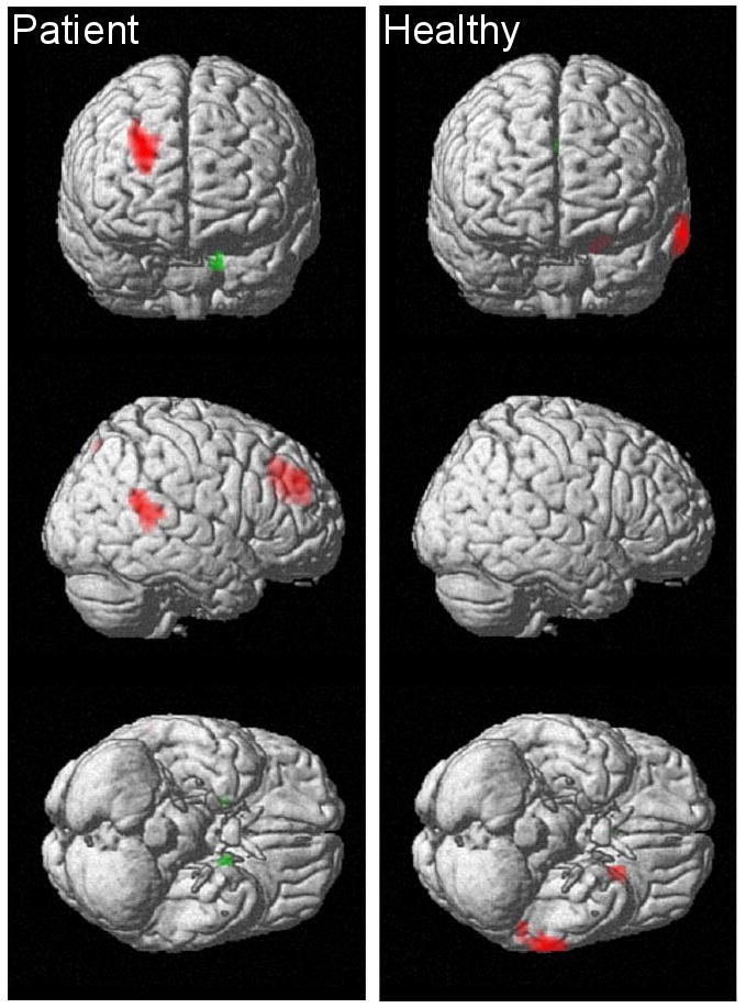
Figure 1.

Statistical PET maps showing areas of significant correlation between COMT genotype and mean resting regional cerebral blood flow (rCBF) in twenty-five, medication-free patients with schizophrenia (right images) and forty-seven healthy volunteers (left images). Red indicates a correlation with the number of met alleles carried and green indicates a correlation with the number of val alleles carried. All regions shown meet an uncorrected voxel-wise height threshold of p<0.001 and extent threshold of 10 voxels. Additionally, neocortical regions meet a cluster-level correction of p<0.05. The right frontal, right superior temporal, precuneus, and left parahippocampal regions indicated in patients also showed significant diagnostic group interactions, indicating the absence of these COMT effects in healthy participants. None of the regions indicated in healthy volunteers showed significant between-diagnostic group differences.
