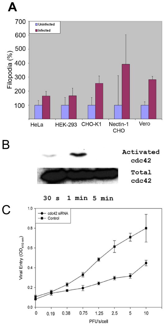
Figure 1. Exogenous HSV-1 can induce filopodia formation.
(A) Increase in the number of filopodia upon exposure to HSV-1 were counted for cells indicated. Counting was done 15–30 min before and after the addition of virus. Numbers are represented as percentages to better compare different cell lines. 3 independent experiments were counted for each cell type (n=25). Numbers of filopodia are presented as means with error bars showing standard deviation. (B) Western Blot analysis confirms activation of Cdc42 at the time points indicated. (C) Down-regulation of Cdc42 inhibits HSV-1 entry. HeLa cells were transfected with siRNA against Cdc42 or control siRNA and exposed to a β-galactosidase expressing HSV-1(KOS) virus.

