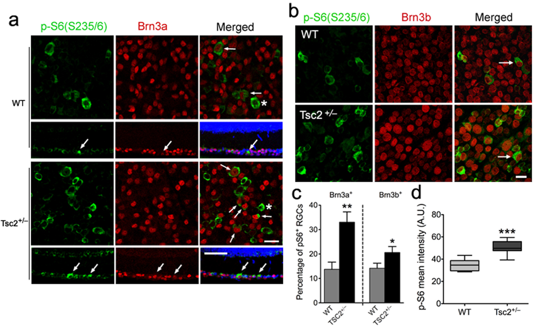
Figure 1. Increased mTOR activity in Tsc2+/− retina in vivo.
(a) Flat mount (first and third row) or horizontal sections (second and fourth row) of retinas of Tsc2+/− and wild-type littermates on postnatal day 23 (P23) were co-stained with antibodies against phospho-S6 (Ser235/236, green) and Brn3a (red). Blue represents nuclear DAPI staining. Arrows denote double-labeled RGCs and asterisks indicate phospho-S6 positive, but Brn3a-negative cells. Scale bars, 25 ⎧m in flat mounts and 50 ⎧m in cross-sections. (b) Flat mount retinas of Tsc2+/− and wild-type littermates (P23) were stained with antibodies against phospho-S6 (Ser235/236, green) and Brn3b (red). Arrows denote phospho-S6 and Brn3b double-labeled RGCs. Scale bar, 20 ⎧m. (c) Quantification of the phospho-S6 positive RGCs as shown in (a) and (b). In both Brn3a- and Brn3b-positive populations, numbers of phospho-S6+ RGCs are significantly increased in Tsc2+/− compared to wild-type littermates (**P < 0.01 and *P < 0.05 by Mann-Whitney tests). (d) Quantification of phospho-S6 (Ser235/236) fluorescence intensity as shown in (a) and (b) from Tsc2+/− versus wild-type (WT) mouse retinas. There was a significant increase in phospho-S6 fluorescence intensity in Tsc2+/− RGCs versus wild-type cells. Data are expressed as mean ± s.e.m. (WT: 34.80 ± 1.909, n = 44 RGCs; Tsc2+/−: 50.76 ± 1.343, n = 114 RGCs; *** P < 0.0001, unpaired t test). A.U. = arbitrary units.

