
Figure 8. Regulation of TSC2 (Ser664) phosphorylation by ephrin-A1 is involved in growth cone dynamics.
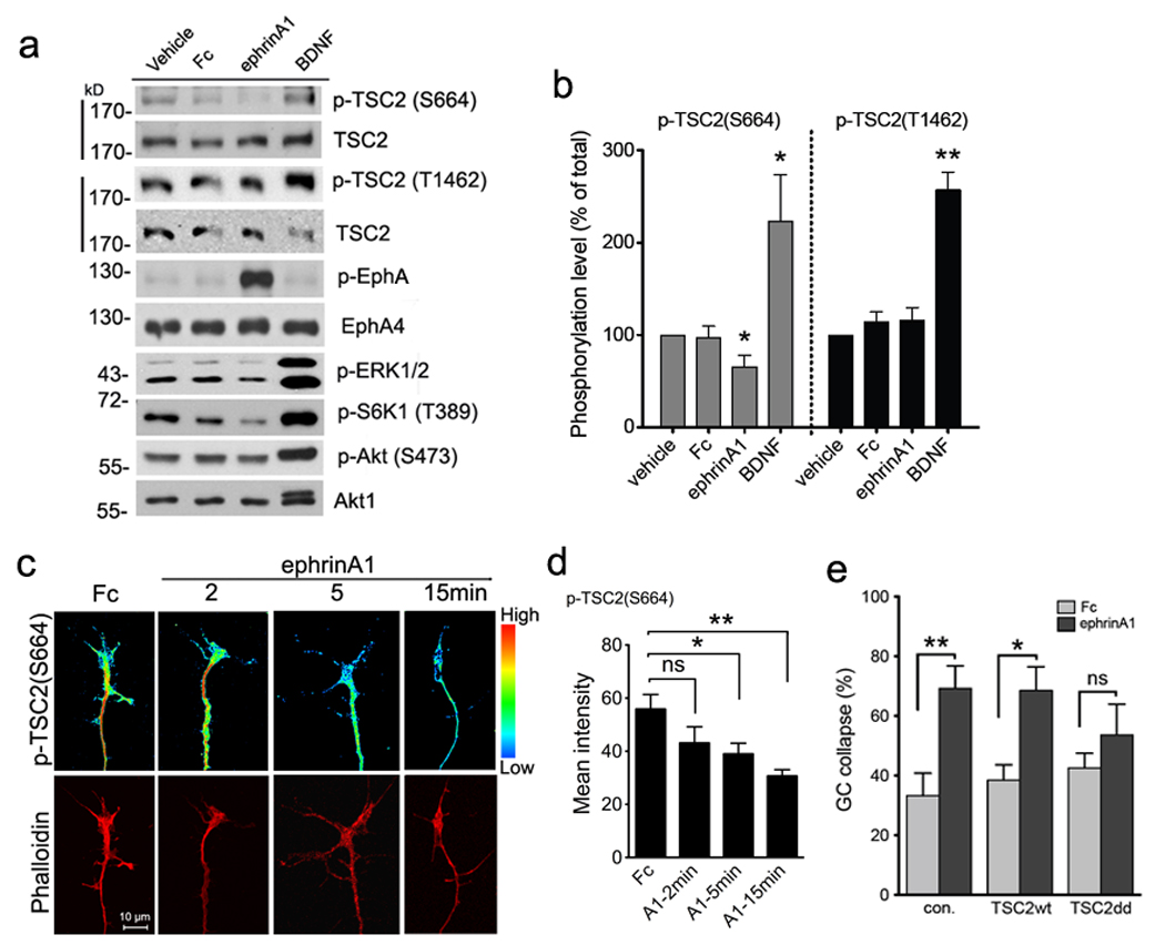
(a) Tsc2+/+ cortical neurons maintained in culture for 12 days were stimulated as indicated for 30 minutes. Phospho-TSC2 (Ser664) or phospho-TSC2 (Thr1462) along with total TSC2 was probed on the same blots and total Akt1 used as the loading control. Full-length gels are presented in Supplementary Figure S14. (b) Intensities of phospho-TSC2 (Ser664) and phospho-TSC2 (Thr1462) were normalized to total TSC2. Data are expressed as % of vehicle-treated intensity ± s.e.m. (* P < 0.05 comparing p-TSC2 (Ser664) in ephrin-A1 versus vehicle, or BDNF versus vehicle; ** P < 0.01 comparing phospho-TSC2 (Thr1462) in BDNF versus vehicle treatment, t-test). (c) Purified RGC cultures from P7 rats were stimulated as indicated and then stained with anti-phospho-TSC2 (Ser664) and rhodamine-phalloidin. Phospho-TSC2 (Ser664) staining was converted to pseudo-colored spectrum display to distinguish intensity change. Scale bar 10 µm. (d) Quantification of pixel intensity of phospho-TSC2 (Ser664) labeling in RGC growth cones. Data represent mean ± s.e.m. (n = 4 – 10 growth cones; * P < 0.05 and ** P < 0.01 and ns =not significant by t-test). (e) Transfection of neurons with phospho-mimic mutant of TSC2 partially blocked growth cone collapse. Rat hippocampal neurons transfected with wild-type TSC2, mutant TSC2 (S664D/S540D) or empty control plasmids were stimulated with Fc or ephrin-A1-Fc for 30 min. Growth cone collapse was determined by phalloidin staining. Data represent mean % collapse ± SEM from 3 independent experiments with duplicates in each experiment (* P < 0.05, ** P < 0.01 and ns =not significant, by t-test).