
Figure 4. Late-stage melanocyte development in larval melanocyte regeneration mutants.
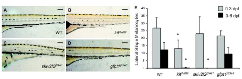
Lateral trunk view at 6 dpf of (A) wild type, (B) kitj1e99, (C) skiv2l2j24e1 and (D) gfpt1j23e1. Scale bars 100 um. (E) Quantitation of embryonic lateral stripe melanocytes at 3 dpf (light gray) and melanocytes that develop from 3–6 dpf (dark gray) in these mutants. Mean value (N=10 for wild type, kitj1e99 and gfpt1j23e1, N=4 for skiv2l2j24e1) with error bars representing standard deviation. * P-values ≤ 0.05.