Figure 3.
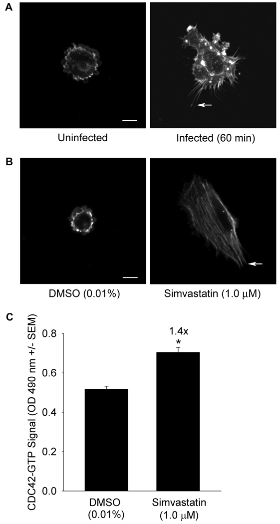
Panel A S. aureus invasion stimulates filopodia formation. Human umbilical vein endothelial cells (HUVEC) were serum starved (20 h) then infected with S. aureus that had been pre-incubated with fibronectin (60 min, 37°C, 5% CO2). Cells were fixed, permeabilized, blocked, and actin detected using Alexa Fluor 488 phalloidin. Arrows indicate representative filopodia. Size bar: 10 µm. Induction was scored in 100 cells/treatment from randomly selected fields. Filopodia formation was greater in infected cells (56%) than in uninfected cells (9%, p ≤ 0.001 by Χ2 test of association). Data and images are representative of replicate experiments. Panel B Simvastatin stimulates filopodia formation. HUVEC were serum starved in the presence of dimethyl sulfoxide (DMSO, 0.01%) or simvastatin (1.0 µM, 20 h, 37°C, 5% CO2). Filopodia were detected and quantified as in Panel A. Filopodia formation was more frequent in simvastatin treated cells (89%) compared to DMSO treated cells (6%, p ≤ 0.001 by Χ2 test of association). Panel C Simvastatin stimulates GTP-loading of CDC42. HUVEC were treated as in Panel B and GTP-bound CDC42, indicated by absorbance at 490 nm, measured by G-LISA. Data are representative of replicate experiments with n = 4/treatment (*significantly greater than DMSO, Student’s t-test, p ≤ 0.05).

