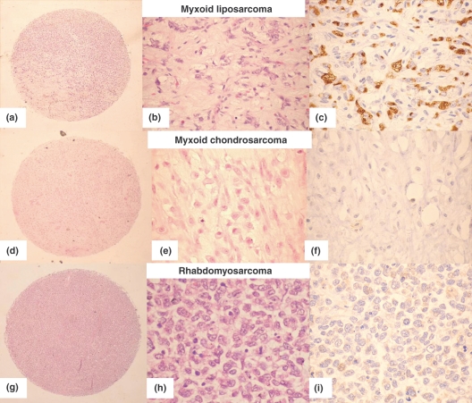
Figure 1.
PIM-1 staining in the TMA. (a–c) Myxoid liposarcoma (H &E, low and high power and PIM-1 showing strong vacuolar cytoplasmic staining). (d–f) Myxoid chondrosarcoma (H&E, low and high power and PIM-1 showing negative staining). (g–i) Rhabdmyosarcoma (H&E, low and high power and PIM-1 showing very pale cytoplasmic non-vacuolar staining).

