Figure 1.

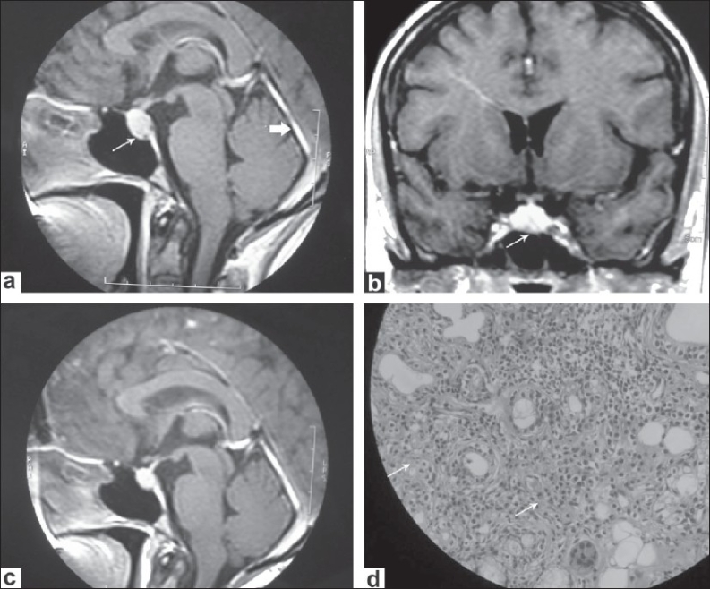
(a) Sagittal gadolinium-enhanced T1-weighted MR images showing thickening and enhancement of cerebellar tentorium (short thick arrow). It also shows a mildly enlarged pituitary gland. (b) Coronal gadolinium-enhanced T1-weighted MR images showing the enlarged and enhancing pituitary gland. (c) Sagittal T1-weighted MR image (post contrast) showing reduction in size of pituitary gland swelling and dural thickening following immunotherapy. (d) Histopathology section of lower lip biopsy showing diffuse infiltration of lymphocytes and plasma cells
