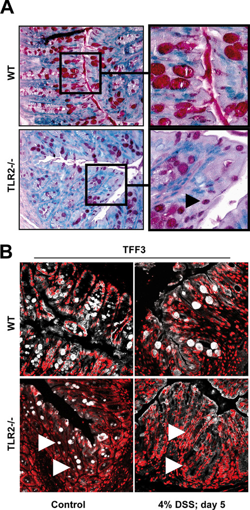
Figure 4. Intestinal TLR2-deficient GC are hypotrophic due to TFF3-deficiency.
(A) Representative PAS-histology of the distal colon of healthy WT [C57BL6/J] or TLR2−/− mice (10x or 40x (insert)). Black arrow indicates example of hypotrophic GC. (B) Representative TFF3 (white) / PI (red) – immunofluorescence of the distal WT or TLR2-deficient colon with or without DSS exposure (4% for 5 days), as assessed by confocal laser microscopy (40x/1.3, oil, scan zoom 0.7). White arrows indicate examples of TFF3-deficient GC.

