Figure 3.
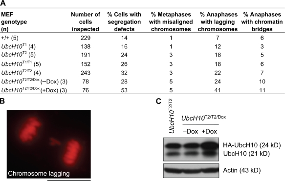
Chromosome missegregation increases as UbcH10 levels rise. (A) Analysis of chromosome segregation defects in MEFs with increasing amounts of exogenous UbcH10. Cells scored as metaphases with misaligned chromosomes displayed congression failure at anaphase onset. UbcH10T2/T2/Dox (+Dox) MEFs were grown in medium containing 1 µg/ml Dox for 2 d before live cell imaging. (B) Image of a transgenic MEF with chromosome lagging. (C) Immunoblots of asynchronous UbcH10T2/T2/Dox MEFs cultured in the presence (+Dox) or absence (−Dox) of 1 µg/ml Dox for 2 d. Blots were probed for UbcH10 and actin. Bar, 10 µm.

