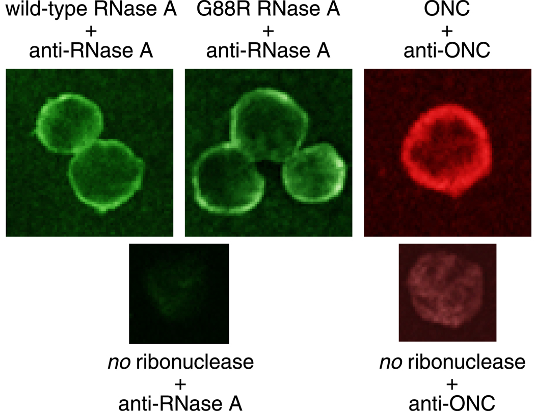
Fig. 2.
Binding of unlabeled RNase A, G88R RNase A, and ONC to the surface of K-562 cells. Cells were incubated with a ribonuclease (1 µM) for 30 minutes at 4°C. Cells were then washed, fixed, and processed for indirect immunofluorescence with antibodies generated against either RNase A or ONC. The appropriate FITC or TRITC-conjugated secondary antibody was used to visualize RNase A (green), G88R RNase A (green), and ONC (red) binding. Negative control samples were incubated in PBS in the absence of protein and processed with primary and secondary antibodies as described above.

